quarta-feira, 6 de dezembro de 2017

Macrófagos


Células redondas de diâmetro e aspetos variáveis (cerca de 16 a 23 µm para os exemplos apresentados). Contêm um ou mais núcleos, nem sempre visíveis, grânulos, vacúolos e outras partículas dispersos no citoplasma.
Morfológicamente poderão ser classificados nos seguintes tipos:
  • ·         Granulares  - citoplasma com grande quantidade de grânulos (3, 4)
  • ·         Vacuolares – citoplasma com número variável de vacúolos (2, 5, 8, 9)
  • ·         Macrófagos com atividade fagocítica –  (1?, 6?)
  • ·         Macrófagos de aspeto homogéneo – citoplasma não contém grânulos ou outras partículas
  • ·        Corpos ovais de gordura (“oval fat bodies”) - macrófagos com citoplasma repleto de gotículas lipídicas.
Quadrado A - Macrófago grânulo-vacuolar binucleado.

O número de macrófagos na urina encontra-se aumentado na nefropatia por IgA e correlaciona-se significativamente com o indíce de atividade histológica na biópsia renal, excreção urinária de leucócitos e rácio proteínas/creatinina na urina da manhã. No entanto, nem sempre é possível justificar a sua presença no sedimento urinário.

Foto IRIS iQ200


quarta-feira, 29 de novembro de 2017

Blastocystis hominis

O ciclo de vida do Blastocystis hominis ainda é alvo de discussão, mas acredita-se que ele alcance o meio exterior através das fezes de indivíduos e animais parasitados e a partir daí se propague para os diversos hospedeiros. Possivelmente a infecção por este parasita ocorre por via fecal-oral, através dos alimentos e águas contaminadas. Quando ingerido, aloja-se no intestino grosso, e é dúbio o seu potencial patogénico. Em alguns casos a infecção será sintomática, enquanto noutros é assintomática. Os principais sintomas relatados são diarreia, cólica, desconforto abdominal, náuseas e vômitos, que podem persistir por semanas ou meses, se não houver tratamento (METRONIDAZOL).

Já foi demonstrada a associação de sintomas com a infecção por este parasita, principalmente em indivíduos imunocomprometidos e transplantados bem como no síndrome do colón irritável.

A forma clássica encontrada nas fezes humanas é o cisto, que pode variar no tamanho entre 5-32  μm. Supõe-se que o cisto de parede espessa presente nas fezes seja o  responsável pela transmissão externa, possivelmente por via fecal-oral através da ingestão de água ou alimentos contaminados. Os cistos infetam células epiteliais do trato digestivo e multiplicam-se assexuadamente. As formas vacuolares do parasita dão origem às multi-vacuolares e às formas amebóides. O multi-vacuolar desenvolve-se em um pré-cisto que dá origem a um cisto de parede espessa considerado responsável pela auto-infeção. A forma amebóide dá origem a um pré-cisto que se desenvolve em cisto de parede espessa por esquizogonia. O cisto de parede espessa é excretado nas fezes.

Estadios de desenvolvimento de Blastocystis hominis (adaptado de  Tan SW, Singh M, Yap EH, et al. (1996). "Colony formation of Blastocystis hominis in soft agar".


Cisto vacuolar

Cisto vacuolar
coloração pelo lugol
Cisto em fissão binária
coloração pelo lugol

Cisto multivacuolar
coloração pelo lugol
 
Forma amebóide
 

Cisto de parede espessa

                                                   © Microbiologia Unilabs Portugal, 2017 (Fotos por P. Laranjeira)

quinta-feira, 27 de abril de 2017

My answers to the CDC DPDx Monthly Case Studies for April 2017 (441 and 442)

Case 441             


A patient, who lives in a rural region in South America, presented with bloody diarrhea and remembers having eaten undercooked pork.  The attending physician submitted images to the DPDx Team of a roundworm he observed during a colonoscopy. Based on the inner structures observed which appeared to be stichocytes, Trichinella spp. was initially suspected as the causative agent. However, an ELISA test for trichinellosis was negative. Figures A-C show what was observed from the colonoscopy. What is your diagnosis?  Based on what criteria?

This case and images were kindly provided by The University of Buenos Aires, University Hospital, Parasitology Division, Buenos Aires, Argentina.

Answer:  Whipworm (Trichuris trichiura) is contracted by ingesting the embryonated eggs 15 to 30 days after being shed and passed with the stool (soil contaminated hands  or food). It is the third most common roundworm to infect humans.


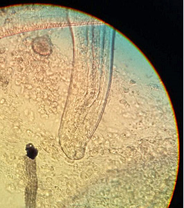
441_A - The mouth is a simple opening without lips and the oesophagus is thin, tubular and surrounded by glandular stichocytes (whole structure referred to as stichosome pharynx).
There are usually no symptoms. Although, severe infections may cause sporadic stomach pains, bloody stools, diarrhea, and weight loss.


441_B - Adult worms have elongate whip-like bodies (3-7cm long), with a long thin anterior end that suddenly becomes thick at the posterior end and live in the cecum and ascending colon.


The adult worms are fixed in that location, with the anterior portions threaded into the mucosa.


441_C- Diverticulum (?);  colonoscopy artifact (?)


Case 442

A 6-year-old child from a community in rural southeastern United States presented with severe upper right abdominal pain, fever, chills, decreased appetite and weight loss.  The child expired 2 weeks after being admitted for symptoms that included hepatitis. At autopsy, a liver biopsy was obtained and processed. Figures A and B show what was observed on a hematoxylin and eosin (H & E) stained liver tissue section at 100x and 500x oil magnification respectively.  What is your diagnosis?  Based on what criteria?

Answer:  C. hepatica causes a serious liver disorder - Hepatic capillariasis.The nematode wanders through the host liver causing tissue damage. The immune response leads to chronic inflammation and encapsulation of the worms in collagen fibers and consequently septal fibrosis (abnormal connective tissue growth) and cirrhosis of the liver. Infections of C. hepatica can present with several clinical symptoms including, abdominal pain in the liver area, weight loss, decreased appetite, fever and chills, hepatitis (liver inflammation), ascites (excess fluid in the peritoneal cavity) and hepatolithiasis (gallstones in the bile ducts).


442_A - Eggs become encased by granulomatous tissue, with large sections of the parenchyma becoming replaced by these egg masses.


442_B - Key identification features of this parasite are a striated shell and shallow polar prominences of the egg.

Capillaria hepatica is rarely found in humans There are no endemic areas of infection with C. hepatica and human infection primarily results from Zoonotic transmission. Of the human infections, most have been found in children under the age of 5.

This parasite can be fatal in humans, as transmission and survival of the parasite depend on death of the definitive host in order for the eggs to reach soil and water to embryonate.

sábado, 25 de março de 2017

Epitélio de Transição - Bexiga


As células redondas e ovais possuem citoplasma abundante e podem exibir pleomorfismo nuclear ou multinucleação e por vezes uma cratera peri-nuclear.

As células basais são pequenas células próximas à membrana basal.
As células basais são encontradas na urina em variadas circunstâncias de lesão do uroepitélio em toda a sua espessura como urolitíase , carcinoma da bexiga ou hidronefrose (doenças urológicas mais frequentes na prática clínica).
Também são frequentes em pacientes com stents ureterais ou cateteres uretrais como consequência da raspagem destes dispositivos na mucosa uroepitelial.


domingo, 26 de fevereiro de 2017

Demodex folliculorum e blefarite crónica

Poster apresentado nas

XX Jornadas Científicas de Análises Clínicas e de Genética Humana
decorrido no dia 8 de outubro de 2016 na Faculdade de Farmácia da Universidade do Porto


My answers to the CDC DPDx Monthly Case Studies for February - 2017 (437 and 438)


Case 437
A 31-year-old female, with travel to Mexico a few months prior, noticed a small worm-like object on the inner surface of her lower lip.  She removed the object with a needle and delivered it to her health care provider.  The object was forwarded to the CDC DPDx Team for identification.  Figures A and B were taken with a dissecting microscope and the ruler in the background shows one centimeter; Figure C is a composite of two images of the organism that was submitted taken with a compound microscope to show the entire organism at 40x magnification.
The insert (of the anterior end) was taken at 100x magnification.  What is your diagnosis?  Based on what criteria?

  Acknowledgements: This case was kindly provided by Optimus Medical Clinic, Houston, Texas.
 
Answer: Gongylonemiasis is the affliction caused by this parasite, which is simply protracted discomfort or sensation of movement in the buccal, oral or gingival areas associated with a sensation of foreign body.

 
Subjects commonly pull worms from their gums, tongue, lips, and inner cheeks after days and even weeks of reported discomfort.

Gongylonema pulchrum is the only parasite of the genus Gongylonema capable of infecting humans. Transmission to humans is due mostly to accidental ingestion of infected coprophagous insects contaminating  food  if unsanitary conditions pervade in its production chain.

The infection usually occurs when someone drinks contaminated water, or consumes an infected beetle.
In humans, there can be an up to six week incubation period for worm development and symptoms may not appear until the second molting of the worm, in which the young adult worms begin migration from the esophagus to the buccal and oral palate tissue. It is this movement through the mucosa of the mouth and lips that causes patients to complain of symptoms. The buccal mucosa with non-keratinized stratified squamous epithelium is the ideal environment for the parasite such as the mucous membrane of the inside of the cheek  contiguous with the mucosae of the soft palate, the undersurface of the tongue and the floor of the mouth.
The average length for male worms is 29.1 mm (1.15 in), while the average length for adult females is 58.7 mm (2.31 in).
The anterior end is covered by numerous cuticular platelets and the buccal opening is small.


Case 438
Fig. A
A 36-year-old male immigrant, with an elevated eosinophil count, was suspected of being infected with pinworms and was administered a pinworm paddle test.  Figures A-B show what was observed on the plastic paddle.  What is your diagnosis?  Based on what criteria?
Acknowledgments: This case and images were kindly provided by the Cadham Provincial Public Health Laboratory, University of Manitoba, Winnipeg, Canada.


Answer: Taeniasis occurs everywhere whenever raw or uncooked beef and/or pork meat infected with encysted  larval stage  are eaten.
Fig. B
Increased eosinophils may be a sign of infection.
Diagnosis of taeniasis is done mainly using stool sample, particularly by identifying the eggs. However, this has limitation at the species level because tapeworms basically have similar eggs.
One possible laboratory diagnostic method is the cellophane-tape technique or a paddle test.


P. Laranjeira (26/02/2017)


Leucócitos na urina


Uma característica dos neutrófilos ativados é a sua capacidade de adesão. Esta característica é essencial para a migração da célula e por isso os neutrófilos podem agregar-se facilmente.


Leucócitos agregados (foto IRIS iQ200)
 
É importante não confundir a agregação de células e pús. O pús é formado de neutrófilos degenerados (piócitos) e restos celulares compactado, numa massa de células onde a identidade é perdida. Esta discriminação não é comumente reportada no sedimento urinário, pelo que que muitos agregados são referidos como pus. O termo “pús” deve restringir-se ao que é ilustrado na figura correspondente.
Piócitos (pús) (foto IRIS iQ200)


P. Laranjeira (26/02/2017)